
Today I was talking about the structure of DNA.
In one part, we were looking at the X-ray diffraction pattern of DNA double helix.
With reference to this webpage, I decided to run a live simulation to mimic what was done by Rosalind Franklin.
It was very simple. I illumined the spring coil (took from a pen) with a laser pointer and pointed it to the screen about 5-7 m away.
A student volunteered to become my scientific assistant and shot the picture below!
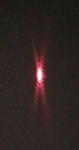
Immediately, this picture was sent by Whatsapp and each student can look at the image simultaneously.
It was cool and the students liked it very much. More importantly, this is going to leave a deep impression in their minds and stimulate their interest in biochemistry.
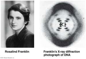
Above is the original image obtained by Franklin and Wilkins for comparison.
Interesting!!
It was Raymond Gosling who took photograph 51. See https://www.nature.com/news/due-credit-1.12806 and https://en.wikipedia.org/wiki/Raymond_Gosling
The DNA model of Watson and Crick is wrong: the two chains are not intertwined but go in parallel. For more details, see my article “DNA: the Double Helix or the Ribbon Helix?” http://vixra.org/abs/1803.0104
Attention: my model radically differs from a well-known Side by Side model.